
Fig. 5
- ID
- ZDB-FIG-250410-32
- Publication
- Lotter et al., 2025 - Dual targeted lipid nanoparticles for enhanced DNA delivery and transfection of breast cancer cells
- Other Figures
- All Figure Page
- Back to All Figure Page
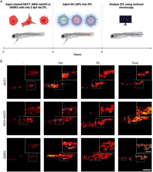
In vivo targeting efficiency of LNPs in a xenograft zebrafish larvae (ZFL) model. A) Schematic representation of the experimental setup to examine the targeting efficiency of targeted LNPs in vivo. Breast cancer cells − MCF7 (low HER2 expression), MDA-mb453 (intermediate HER2 expression), and SKBR3 (strong HER2 expression) − were stained with CellTracker Deep Red stain and injected into 2 days post-fertilization (dpf) old ZFL to reach a final concentration of 250 cells per ZFL. 3 hp cell injection ZFL were further treated with DiI-labeled LNPs (Her-, FA-, and Dual-LNPs; 0.75 pg encapsulated DNA/ZFL) and imaged 1 hp LNP injection. B) Representative confocal images of tail region of ZFL, showing breast cancer cells (red) and DiI-labeled LNPs (blue). Overlap of MCF7, MDA-mb453, and SKBR3 cells with DiI-labeled LNPs is colored in yellow. Insert: magnified section of dotted area. Results are shown for control ZFL and Her-, FA-, and Dual-LNP treated ZFL 3 hp cell and 1 hp LNP injection. Scale bar = 100 µm. Scale bar inserts = 50 µm. (For interpretation of the references to colour in this figure legend, the reader is referred to the web version of this article.) |