
Fig. 7
- ID
- ZDB-FIG-250410-13
- Publication
- Ramkumar et al., 2025 - Phased ERK responsiveness and developmental robustness regulate teleost skin morphogenesis
- Other Figures
- All Figure Page
- Back to All Figure Page
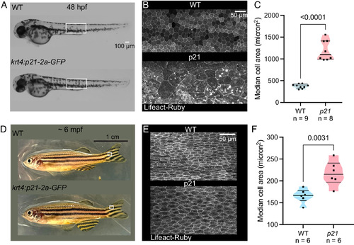
Periderm growth exhibits adaptive robustness. (A) Brightfield view of WT control and krt4:p21-2a-GFP embryos at 48 hpf, showing normal axial elongation. (Scale bar, 100 µm.) (B) Maximum projection view of periderm cells in the trunk region of representative larvae boxed in A expressing Lifeact-Ruby to outline cell boundaries. Periderm cell area is massively increased in embryos expressing p21. Asterisk, abnormal accumulation of actin in periderm cells. (Scale bar, 50 µm.) (C) Median cell area of control and p21-expressing embryos at 48 hpf. Average cell size in control embryos at 48 hpf, 373.1 micron2, n = 9 larvae; p21-expressing embryos, 1,195 micron2, n = 8 larvae. P < 0.0001, unpaired t test. (D) Brightfield view of WT and p21-expressing adult fish at 6 months, showing normal morphology. (Scale bar, 1 cm.) (E) Maximum projection view of periderm cells in the fin region of representative adult fish boxed in D expressing Lifeact-Ruby to outline cell boundaries. Periderm cell area is slightly increased in p21-expressing adults. (Scale bar, 50 µm.) (F) Median cell size of periderm cells covering fin region quantified in control and p21-expressing adults. Average cell area in control fish, 165.7 micron2, n = 6 adult fish; p21-expressing fish, 217.2 micron2, n = 6 adult fish. P = 0.0031, unpaired t test. |