
Fig. 1
- ID
- ZDB-FIG-250407-18
- Publication
- Chen et al., 2025 - Loss-of-Function of CLMP Is Associated With Congenital Short Bowel Syndrome and Impaired Intestinal Development
- Other Figures
- All Figure Page
- Back to All Figure Page
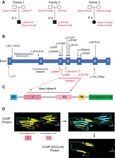
Genetic analysis of CSBS patients. (A) Pedigree of the three families in this study with CLMP genetic mutations. (B) Schematic overview of all the variants identified in the human CLMP gene that coincide with CSBS. In red are the variants reported in our center and in black are the variants previously described. Numbering starts with position 1 at the amino-terminal methionine of the unprocessed polypeptide. The single letter code for amino acids is used, with one asterisk (*) indicating translation stop. (C) Schematic diagram of human CLMP protein structures. C2, C2-typed Ig-loops; SS, signal sequence; TM, transmembrane region; V, V-typed Ig-loops. (D) Crystal structure of the extracellular region of CLMP protein tertiary structure was calculated by Phyre2 based on CAR (PDB: 3jz7A) as the template for molecular modeling. The CLMP Model dimensions (Å): X:60.895 Y:40.957 Z:82.390. The asterisks mark the amino-terminal and carboxyl-terminal. The blue region represents the corresponding amino acid sequence of exons 3–5. CAR, coxsackievirus-adenovirus receptor. |