FIGURE

FIGURE 7

ID
ZDB-FIG-250320-28
Publication
Jeon et al., 2025 - Pax1a-EphrinB2a pathway in the first pharyngeal pouch controls hyomandibular plate formation by promoting chondrocyte formation in zebrafish
Other Figures
All Figure Page
Back to All Figure Page
FIGURE 7

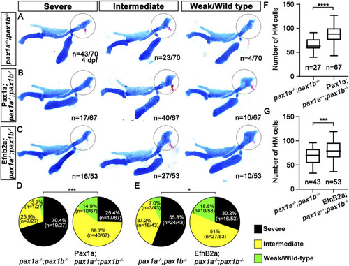
Requirement of Pax1a-EphrinB2a in the first pouch for hyomandibular plate development. (A–C) Unilateral dissections of the skeletons of the mandibular and hyoid arches stained with Alcian Blue (cartilage) and Alizarin Red (bone) at 4 dpf. HM plates are circled. The severe, intermediate, and weak defects in HM are seen in both pax1 double mutants (A) and their siblings expressing Pax1a (B) or EfnB2a (C) in sox17-positive endoderm. Anterior is to the left. (D, E) Quantification of the frequency of each group between pax1 double mutants and their siblings carrying Tg(sox17:Gal4VP16) and Tg(UAS:Pax1a)(D) or Tg(sox17:Gal4VP16) and Tg(UAS:EfnB2a) transgenes (E). The frequency of each group is counted. Data is represented on a pie chart. Black, yellow, and green represent the severe, intermediate, and weak/wild-type groups. *** and * indicate p < 0.001 and p < 0.05, respectively. (F, G) Quantification of the number of cells in HM. The number of cells in HM is counted. Data is represented on a boxplot. **** and *** show p < 0.0001 and p < 0.001, respectively. n, number of animals analyzed.

Expression Data

Expression Detail
Antibody Labeling
Phenotype Data

Phenotype Detail
Acknowledgments
This image is the copyrighted work of the attributed author or publisher, and ZFIN has permission only to display this image to its users. Additional permissions should be obtained from the applicable author or publisher of the image. Full text @ Front Cell Dev Biol