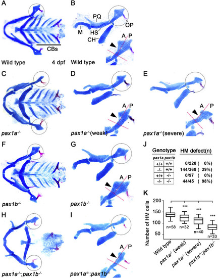
Requirement of pax1a in hyomandibular cartilage development. (A–I) Craniofacial skeletal elements stained with Alcian Blue (cartilage) and Alizarin Red (bone) at 4 dpf, with the hyomandibular (HM) plate circled. (A, C, F, H) Ventral views of dissected facial cartilages. (B, D, E, G, I) Lateral views of dissected facial skeletal elements of the mandibular and hyoid arches, along with inserts showing the anterior and posterior regions of HM (circled areas). (A, B) All facial skeletal elements are present in wild types, including the triangular HM plate, with an arrowhead indicating the multi-layered cells in the anterior region of HM. Five CBs on one side are underlined. (C–E) In pax1a mutants, a specific defect is seen in the HM plate, with other skeletal elements unaffected. Arrowheads indicate mono-layered cells (D) and missing cells (E) in the anterior region of HM. (F, G) In pax1b mutants, all facial skeletal elements are normal, including the HM plate. The normal multi-layered cells in the anterior region of HM are marked with an arrowhead (H, I) In double mutants for pax1a and pax1b, the HM plate is defective, and most CB cartilages are missing. Arrowhead indicates the missing cells in the anterior region of HM. M, Meckel’s cartilage; PQ, Palatoquadrate cartilage; HS, Hyosymplectic cartilage; CH, Ceratohyal cartilage; CB, Ceratobranchial cartilage; OP, Opercle bone; A, anterior; P, posterior. Anterior is to the left. (J) The number of mutants analyzed and the penetrance for HM defects. (K) Quantification of the number of cells in HM. Data is represented on a boxplot. *** shows p < 0.001.
|
