|
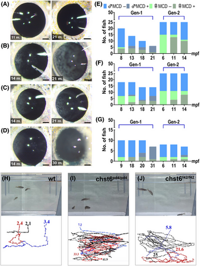
chst6 mutant zebrafish develop MCD and impaired vision. Macroscopic analysis of (A) wt, (B) chst6pd4/pd4, (C) chst6pd5/pd5, and (D) chst6f42/f42 alleles. Eyes of young (11–14 mpf) adults of gen2 (left), old (21–30 mpf) adults of gen1 (right) are shown. Scale bar: 300 μm. (E–G) Incidence of opaque corneal aggregates homozygous mutants of two generations over time (E) Green bar: females, blue bar: males, dashed bars indicate occurrence of opaque aggregates. (E) chst6pd4/pd4, (F) chst6pd5/pd5, and (G) chst6f42/f42. (H–J) The vision test was performed with wt and homozygous chst6 mutants. Swim paths and durations were tracked. Track was ended if the fish reached food and time stamp is displayed at the end of each track. (H) wt fish swam for 2.4 and 3.4 s until it reached the food (n = 3). (I) chst6pd4/pd4 swam for 33.3 and 41.9 s without noticing the food (n = 3). (J) chst6f42/f42 swam for 21.6 and 25 s without noticing the food (n = 3).
|