
Fig. 7
- ID
- ZDB-FIG-250210-7
- Publication
- Nurhidayat et al., 2025 - Tokay gecko tail regeneration involves temporally collinear expression of HOXC genes and early expression of satellite cell markers
- Other Figures
- All Figure Page
- Back to All Figure Page
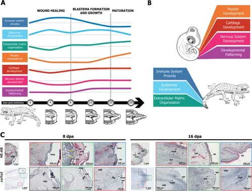
A summary of selected key features of tail regeneration in the tokay gecko. A Schematic overview of biological process enrichment in the transcriptome at selected stages of tail regeneration. The height of each line represents (schematically) the relative enrichment or the process. The greyscale drawings at the bottom represent the histological structure of the regenerating tail at that stage. B Schematic illustration highlighting the major differences in the dominant pathways in the regenerating tail vs. the embryonic tail. C There is no apical growth zone in the regenerating tail of the tokay gecko. The tissues immediately under the wound epithelium (pre-blastema) are dominated by col1a2+ connective tissue cells. Key: adp adipose tissue, amc adult muscle cells, bls blastema, bvl blood vessel, ep epidermis, ery erythrocytes, leu leukocytes, myb myoblast, pbl pre-blastema, scb wound scab, wep wound epithelium |