
Fig. 2
- ID
- ZDB-FIG-250206-13
- Publication
- Chen et al., 2024 - Exposure to Nanoplastics Cause Caudal Vein Plexus Damage and Hematopoietic Dysfunction by Oxidative Stress Response in Zebrafish (Danio rerio)
- Other Figures
- All Figure Page
- Back to All Figure Page
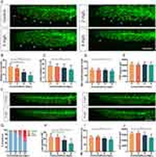
Exposure to NPs leads to developmental defects in the CVP of Tg(kdrl:eGFP) embryos. (A) Representative images of CVP developmental malformations in Tg(kdrl:eGFP) embryos treated with NPs at 30 hpf. (B-E) The numbers of filopodia (B), numbers of loop (C), maximum ventral extension distance (D) and area (E) of CVP in Tg(kdrl:eGFP) embryos after quantified NPs treatment at 30 hpf (n = 30 embryos). (F) Representative images of CVP developmental malformations in NPs-treated Tg(kdrl:eGFP) embryos at 48 hpf. (G) The malformation rate of CVP in Tg(kdrl:eGFP) embryos after quantified NPs treatment at 48 hpf (n = 30 embryos). (H-J) The numbers of loop (H), maximum ventral extension distance (I) and area (J) of CVP in Tg(kdrl:eGFP) embryos after quantified NPs treatment at 48 hpf (n = 30 embryos). White arrowheads, sprouts; red asterisks, loop structure; white vertical line, maximum ventral extension distance. Data are shown as mean ± SD. An asterisk above each bar indicates statistical significance (*P<0.05; **P<0.01; ***P<0.001). Scale bar, 100 μm. |