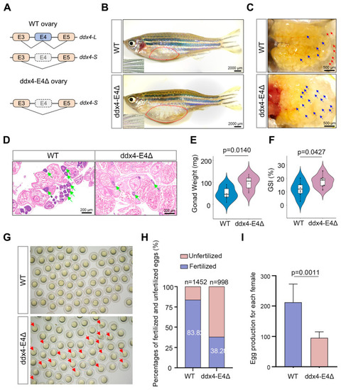
ddx4-L knockout causes reduced fecundity of female zebrafish. (A) Alternative splicing patterns of ddx4 in WT and ddx4-E4Δ zebrafish ovaries are shown. (B) Abdominal morphology and secondary sexual characteristics of WT and ddx4-E4Δ zebrafish at 9 mpf. The insets show the pectoral fin of zebrafish. Red dotted frames indicate the position of ovaries. n = 7. Scale bars: 2000 μm. (C) Overall ovarian morphology of WT and ddx4-E4Δ zebrafish at 9 mpf. The red arrows indicate examples of mature eggs, and the blue arrows indicate examples of immature eggs. n = 7. Scale bars: 500 μm. (D) Hematoxylin and eosin (HE) staining of WT and ddx4-E4Δ ovarian sections at 9 mpf. The green arrows indicate early-stage oocytes, which have large, spherical nuclei stained blue or purple due to the high affinity of hematoxylin. n = 7. Scale bars: 200 μm. (E) Ovarian weights of WT and ddx4-E4Δ zebrafish at 9 mpf are shown. n = 7. (F) GSI of WT and ddx4-E4Δ females at 9 mpf are shown. n = 7. (G) Embryos produced by WT and ddx4-E4Δ zebrafish are shown. The arrows indicate the unfertilized embryos. (H) Fertilization rates of spawning eggs were quantified for WT and ddx4-E4Δ zebrafish. (I) The number of eggs produced by a single female during a spawning cycle was quantified. The ddx4-E4Δ females laid fewer eggs than WT controls. n = 6.
|
