FIGURE

Fig 7

ID
ZDB-FIG-250109-260
Publication
Fietta et al., 2024 - Neuroblastoma-derived hypoxic extracellular vesicles promote metastatic dissemination in a zebrafish model
Other Figures
All Figure Page
Back to All Figure Page
Fig 7

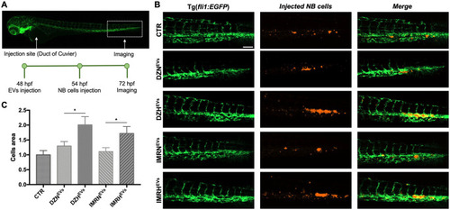
Hypoxic EVs promote the proliferation of NB cells in the CHT.

A. Injection site, analysis site, and timeline for Tg(fli1:EGFP) embryos injected with NB-derived EVs and NB cells. Embryos were injected with EVs isolated from SK-N-DZ or IMR32 cells cultured in normoxic or hypoxic conditions, and subsequently injected with SK-N-DZ and IMR32 cells, respectively. B. Representative CHT images 24 hpi of EVs and 18 hpi of SK-N-DZ cells (orange). Scale bar 100 μm. C. Representative CHT images 24 hpi of EVs and 18 hpi of IMR32 cells (orange). Scale bar 100 μm. D. Evaluation of proliferated SK-N-DZ cells area in the CHT at 72 hpf. n = 53 from at least 3 independent experiments. * p < 0.05. E. Evaluation of proliferated IMR32 cells area in the CHT at 72 hpf. n = 59 from at least 3 independent experiments. * p < 0.05.

Expression Data

Expression Detail
Antibody Labeling
Phenotype Data

Phenotype Detail
Acknowledgments
This image is the copyrighted work of the attributed author or publisher, and ZFIN has permission only to display this image to its users. Additional permissions should be obtained from the applicable author or publisher of the image. Full text @ PLoS One