|
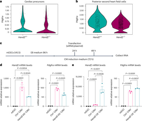
Hand2 promotes Pdgfra expression in mouse cardiac cells. a, Violin plots showing Pdgfra expression in cardiac precursors of E8.25 WT and Hand2−/− mutant hearts (data analyzed from GSE126128 (ref. 56)). b, Violin plots showing Pdgfra expression in posterior second heart field cells of E8.25 WT and Hand2−/− mutant hearts (data analyzed from GSE126128 (ref. 56)). c, Schematic of knockdown and overexpression of Hand2 during mESC differentiation into cardiac cells. d, Relative mRNA levels of Hand2 and Pdgfra in Hand2 knockdown mESC-derived cardiac cells; error bars are mean ± s.e.m.; n = 3 biologically independent samples. e, Relative mRNA levels of Hand2 and Pdgfra in Hand2 OE mESC-derived cardiac cells; error bars are mean ± s.e.m.; n = 3 biologically independent samples. P values in d and e were calculated using a one-way ANOVA multiple-comparison test. The average mRNA level in mESCs was set at 1.0. The Ct values of qPCR data are listed in Supplementary Table 1. Ctrl, control; KD, knockdown.
|