|
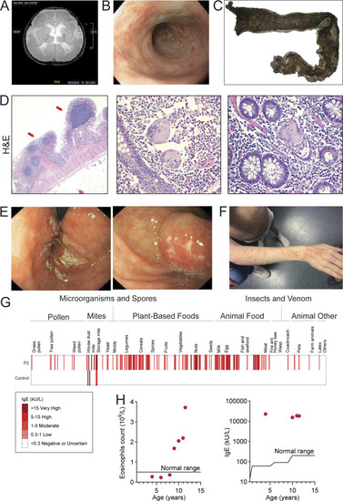
Clinical features. (A and B) Brain MRI (A) and colonoscopy (B) of P1. (C) Gross image showing colectomy specimen from P1 with diffuse pancolitis. (D) H&E stains, left (10× magnification), showing full thickness section of the distal colon with inflammation limited to the superficial submucosa and inflammatory pseudopolyps with lymphoid aggregates (arrows). The uninflamed muscularis propria and subserosa with congested vessels can be seen. (D) Center and right (200× magnification), showing lamina propria with numerous neutrophils and eosinophils accompanying two multinucleated giant cells, indicating a site of crypt rupture. (E) Colonoscopy of P2. (F) Image of P2 showing severe atopic dermatitis. (G) Screening for IgE against 295 allergens with the ALEX Allergy Explorer. (H) Eosinophil counts in whole blood (n = 7), normal upper limit <0.5 × 10⁹/liter; and IgE concentrations (n = 4), normal upper limit (IgE < 100 kU/liter) for P2 (red dots).
|