
Fig. 2
- ID
- ZDB-FIG-240916-188
- Publication
- Roider et al., 2024 - MITF regulates IDH1, NNT, and a transcriptional program protecting melanoma from reactive oxygen species
- Other Figures
- All Figure Page
- Back to All Figure Page
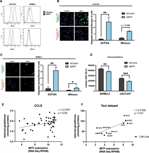
MITF protects melanoma cells from oxidative stress in vitro. (A) Representative flow plots for cytosolic ROS levels measured by DCFDA fluorescent dye and mitochondrial ROS levels measured by Mitosox fluorescent dye in UACC257 melanoma cells after knockdown of MITF. (B) Confocal images of cytosolic ROS levels measured by DCFDA fluorescent dye and mitochondrial ROS levels measured by Mitosox fluorescent dye in UACC257 melanoma cells after knockdown of MITF. Representative images (left panel) and quantification of three independent experiments (right panel, n.s: p = 0.056) are displayed. (C) Confocal images of cytosolic ROS levels measured by DCFDA fluorescent dye and mitochondrial ROS levels measured by Mitosox fluorescent dye in SKMEL30 melanoma cells after knockdown of MITF. Representative images (left panel) and quantification of three independent experiments (right panel) are displayed. (D) Reduced glutathione (GSH) was measured in UACC257 and SKMEL5 melanoma cells after siRNA silencing of MITF. Reduced glutathione levels significantly correlate with MITF expression in melanoma cell lines in CCLE (n = 51, Pearson’s r = 0.32, p < 0.05) (E) and in patient-derived melanoma cells (n = 10, Pearson ‘s r = 0.77, p < 0.01) (F). (Total available melanoma cell lines for microarray data at the Depmap portal: 61) Non-targeting siRNA (siControl) was used as negative control. Values represent the mean ± SEM. *p < 0.05, **p < 0.01, ***p < 0.01. Abbreviation: MITF microphthalmia-associated transcription factor, ROS reactive oxygen species, DCFDA dichlorodihydrofluorescein diacetate, CCLE cancer cell line encyclopedia. |