
Fig. 5
- ID
- ZDB-FIG-240910-63
- Publication
- Dill et al., 2024 - Neuropeptidergic regulation of neuromuscular signaling in larval zebrafish alters swimming behavior and synaptic transmission
- Other Figures
- All Figure Page
- Back to All Figure Page
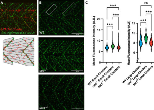
Abundance of nAChRs on muscle cells is altered in neuropeptide mutants (A) Representative α-bungarotoxin staining at 4 dpf (upper panel). nAChR clusters in green, bPAC-expressing motor neurons in red. Diagram of skeletal muscle cells and clusters of nAChRs on the cell surface (lower panel). Large receptor clusters are assembled on somite boundaries; small receptor clusters are distributed in between. (B) α-bungarotoxin staining at 4 dpf on wild-type, cpe−/−, and tac1−/− animals. The rectangle in the upper panel represents an area containing small receptor clusters on fast muscle; the dashed rectangle marks larger clusters at the end of slow muscle cells. (C) Quantification of fluorescence intensity of small and large receptor clusters in the respective zebrafish strains. Median and 25/75 quartiles (thick and thin black lines), min to max are shown. Scale bars in (B): 100 μm. One-way ANOVA with Tukey multiple comparisons of means. Statistical significance given as ∗∗∗p < 0.001; n.s., non-significant. |