
Fig. 8
- ID
- ZDB-FIG-240904-19
- Publication
- Sam et al., 2024 - Gata6 functions in zebrafish endoderm to regulate late differentiating arterial pole cardiogenesis
- Other Figures
- All Figure Page
- Back to All Figure Page
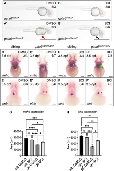
BCI treatment partially rescues cardiac defects in gata6wcm7/wcm7 mutants. (A-B′) Bright-field images of 2 dpf gata6 mutant and sibling embryos exposed to DMSO (A,A′) or 7.5 µM BCI (B,B′) for 6 h starting at the 15-somite stage. Pericardial edema is essentially eliminated in BCI-treated mutants (B′, black arrow) compared with DMSO-treated controls (A′, red arrow). Scale bars: 1000 µm. (C-F′) Whole-mount in situ hybridization (WISH) analysis revealed largely restored expression domains of vmhc (D′) and elnb (F′) in gata6 mutants after overnight exposure to BCI starting at the 15-somite stage (compare C′ and E′, respectively). White bars indicate the normal length of the ventricle or outflow tract in DMSO-treated siblings. Yellow (C′,D′) and red (E′,F′) bars indicate shortened length in the DMSO-treated mutants. Green bars indicate restored length after overnight exposure to BCI. Scale bars: 100 µm. (G,H) Quantification of WISH staining confirmed a statistically significant increase in the area of vmhc and elnb expression domains with BCI treatment (one-way ANOVA, *P<0.05, ***P<0.001 and ****P<0.0001). Data are mean±s.e.m. |