
Fig. 6
- ID
- ZDB-FIG-240814-33
- Publication
- Beaulieu et al., 2024 - Transdifferentiation is temporally uncoupled from progenitor pool expansion during hair cell regeneration in the zebrafish inner ear
- Other Figures
- All Figure Page
- Back to All Figure Page
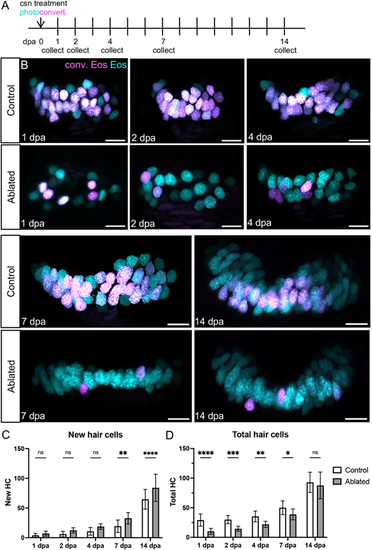
Anterior crista hair cells regenerate during the 2 weeks that follow ablation. (A) Tg(myo6b:NLS-Eos) sibling larvae with or without Tg(myo6b:TrpV1-mClover) were photoconverted and treated with capsaicin to ablate hair cells at 8 dpf. Larvae were collected at five timepoints over the following 2 weeks: 1 (n=22 control, 25 ablated), 2 (n=13, 20), 4 (n=19, 18), 7 (n=16, 13) or 14 (n=18, 15) days-post ablation. (B) Representative maximum intensity projections of anterior crista in control and ablated fish at five timepoints after treatment. Nuclei of cells that survived capsaicin treatment contain photoconverted Eos (magenta). Hair cells newly added after capsaicin treatment have nuclei with only unconverted Eos (cyan). Scale bars: 10 µm. (C) Quantification of new (cyan only) hair cells in ablated and control anterior crista. Two-way ANOVA variation across condition, P<0.0001; Šídák's multiple comparisons post-hoc test for 7 dpa, **adjusted P-value=0.0021, and for 14 dpa, ****adjusted P-value<0.0001. (D) Quantification of total hair cells in ablated and control anterior crista. Two-way ANOVA variation across condition, ****P<0.0001; Šídák's multiple comparisons post-hoc test for 1 dpa, ****adjusted P-value<0.0001, for 2 dpa, ***adjusted P-value=0.0006, for 4 dpa, **adjusted P-value=0.0015, and for 7 dpa *adjusted P-value=0.0342. Data are mean±s.d. |