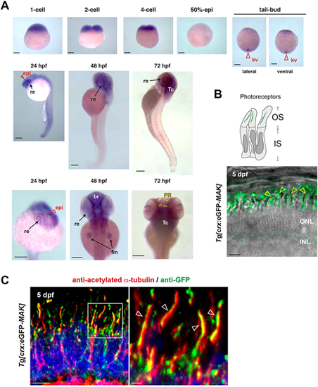
Mak is localized in axonemes of photoreceptor cilia. (A) Whole-mount in situ hybridization of zebrafish embryos with mak RNA probe. mak mRNA expression was observed in Kupffer's vesicles (red open arrowheads) at the tail-bud stage, in the retina (arrows) and epiphysis (red open arrowheads) at 24 hpf, and in the retina, brain and fin (arrows) at 48 hpf. Retinal expression was restricted to the photoreceptor layer (yellow arrowheads) at 72 hpf. kv, Kupffer's vesicle; re, retina; epi, epiphysis; br, brain; fin, pectoral fin; Tc, optic tectum; PR, photoreceptor layer. (B) Confocal live image of Tg[crx: eGFP-MAK] transgenic retina at 5 dpf. eGFP-Mak (green) was localized in the OS of photoreceptors (yellow arrowheads). (C) Labeling of 5-dpf Tg[crx: eGFP-MAK] transgenic retina with anti-acetylated α-tubulin (red) and anti-GFP (green) antibodies. Nuclei were counterstained with Hoechst 33342 (blue). The right panel shows a higher magnification of the square shown in the left panel. eGFP-Mak was localized in the axoneme (white open arrowheads, right panel). Sample sizes are shown in Table S3. Scale bars: 200 µm (A); 5 µm (B, left panel in C); 1 µm (right panel in C).
|
