
Fig. 1
- ID
- ZDB-FIG-240703-97
- Publication
- Lo et al., 2024 - Primary cilia formation requires the Leigh syndrome-associated mitochondrial protein NDUFAF2
- Other Figures
- All Figure Page
- Back to All Figure Page
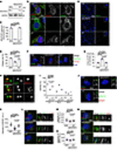
Loss of NDUFAF2 in RPE cells results in primary cilia defects. (A) NDUFAF2WT was stably expressed in NDUFAF2–/– cells. Western blot analysis was performed with antibodies against NDUFAF2 and GAPDH. (B) Cells stained with TOMM20 (green) and NDUFAF2 (red) antibodies. DNA stained with DAPI (blue). Scale bar: 10 μm. (C) NDUFAF2 staining of mitochondria in wild-type, NDUFAF2–/–, and NDUFAF2WT-re-expressing RPE1 cells. (D) Cells stained with polyglutamylated tubulin (green) antibodies. DNA stained with DAPI (blue). Scale bar: 10 μm. (E) Percentage of ciliated cells after serum starving for 2 days; >150 cells analyzed for each independent experiment. (F) Immunofluorescent analysis of cells serum-starved for 2 days. Cells stained with CP110 (red), CEP164 (green), and FOP (FGFR1 oncogene partner; blue) antibodies. DNA stained with DAPI (blue). Scale bars: 10 μm. (G) Graph shows percentage of serum-starved cells with two CP110 dots at the centrioles; >150 cells analyzed for each independent experiment. (H) Immunofluorescent analysis of cells serum-starved for 6 hours. Cells were stained with myosin-Va (red) and centrin (green) antibodies. DNA stained with DAPI (blue). Scale bar: 2 μm. (I) Percentage of cells with ciliary vesicles demonstrated by an antibody against myosin-Va after serum starving for 6 hours (CV, ciliary vesicle; NCV, no ciliary vesicle; PCV, preciliary vesicle); >150 cells analyzed for each independent experiment. (J) Immunostaining of cells serum-starved for 2 days. Scale bars: 10 μm. (K) Quantification of NPHP1 signal intensity at the centrioles; >100 cells analyzed for each independent experiment. (L) Immunofluorescent analysis of cells serum-starved for 2 days. Cells stained with TCTN2 (red) and pGlu-Tu (green) antibodies. DNA stained with DAPI (blue). Scale bars: 10 μm, 2 μm. (M) Quantification of TCTN2 signal intensity at the centrioles; >50 cells analyzed for each independent experiment. (N) Immunofluorescent analysis of cells serum-starved for 2 days. Cells stained with MKS1 (red) and pGlu-Tu (green) antibodies. DNA stained with DAPI (blue). Scale bars: 10 μm, 2 μm. (O) Quantification of MKS1 signal intensity at the centrioles; >50 cells analyzed for each independent experiment. Bars represent mean ± SD, n = 3. Exact P values are indicated. ANOVA followed by Tukey-Kramer multiple-comparison test. |