FIGURE

Figure 7

ID
ZDB-FIG-240702-159
Publication
Song et al., 2024 - The structure of TRAF7 coiled-coil trimer provides insight into its function in zebrafish embryonic development
Other Figures
All Figure Page
Back to All Figure Page
Figure 7

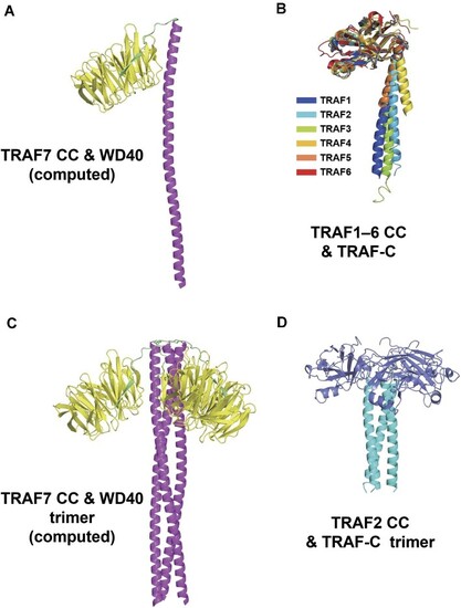
C-terminal structural alignment of TRAF family members. (A) AlphaFold-predicted structure of TRAF7 CC and WD40 domains. The CC region (288–374 aa) is a long α-helix colored magenta, while WD40 (385–670 aa) is colored yellow. (B) Alignment of crystal structures of TRAF1–6 CC and TRAF-C regions. TRAF1–4 and TRAF6 are from humans, and TRAF5 is from mice. TRAF1–5 are homotrimers in crystals, which only show monomers. PDB ID: TRAF1, 5E1T; TRAF2, 1CA9; TRAF3, 1FLK; TRAF4, 3ZJB; TRAF5, 4GJH; TRAF6, 1LB4. (C) TRAF7 CC and WD40 trimer generated by AlphaFold2.3.1 with TRAF7 homotrimer input. (D) TRAF2 CC and TRAF-C trimer from the crystal structure (PDB ID: 1CA9). The CC domain (311–347 aa) is an α-helix colored cyan. The TRAF-C domain (352–501 aa) is colored slate blue.

Expression Data

Expression Detail
Antibody Labeling
Phenotype Data

Phenotype Detail
Acknowledgments
This image is the copyrighted work of the attributed author or publisher, and ZFIN has permission only to display this image to its users. Additional permissions should be obtained from the applicable author or publisher of the image. Full text @ J. Mol. Cell Biol.