
Fig. 6
- ID
- ZDB-FIG-240621-56
- Publication
- Zeitz et al., 2024 - Variants in UBAP1L lead to autosomal recessive rod-cone and cone-rod dystrophy
- Other Figures
- All Figure Page
- Back to All Figure Page
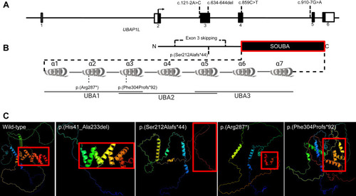
Gene structure, prediction of domains and 3D-modeling of the unaffected and altered UBAP1L protein. A. Schematic representation of UBAP1L gene structure with the positions of identified variants. B. Schematic representation of UBAP1L protein structure with the positions of identified variants. The SOUBA domain structure is detailed showing the composition of UBAs and α-helices. C. AlphaFold 3D structure predictions of UBAP1L wild-type (UniprotKB: F5GYI3), p.(His41_Ala233del), p.(Ser212Alafs∗44), p.(Arg287∗), and p.(Phe304Profs∗92). The color scheme is a gradient that goes from N terminus (blue) to C terminus (red). |