Fig. 3
|
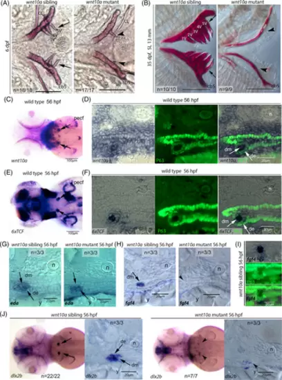
unm_t30922t30922 mutants display impaired tooth development. (A) At 6 dpf, the wnt10a siblings have alizarin red-stained 3V1, 4V1, and 5V1 teeth on either bilateral ceratobranchial 5 arch (cb5), while the unm_t30922t30922 mutant either has one tooth or no teeth on either side of an otherwise normally formed cb5. The cleithra (CL) of the mutant is indistinguishable from those of the siblings. (B) At 35 dpf (SL 13 mm), dissected cb5 of siblings have fully formed alizarin red-stained mineralized teeth (only 1V, 2V, 3V, 4V, and 5V are visible in this view) while the unm_t30922t30922 mutants have no teeth and an underdeveloped cb5. Arrows indicate regular teeth, arrowheads compromised or absent teeth. Scale bars = 100 μm. (C) Dorsal view of wild-type embryo at 56 hpf, after wnt10a in situ hybridization shows that wnt10a is expressed in the tooth germs (indicated by arrows). (D) Transverse cryosection of 56 hpf wild-type embryo after wnt10a in situ hybridization and p63 immunostaining, revealing strong wnt10a expression in the dental mesenchyme (dm; white arrow) and weak wnt10a expression in the surrounding p63-positive dental epithelium (de; white arrow). (E) Dorsal view of Tg(6xTCF:eGFP) embryo at 56 hpf after transgene-encoded GFP in situ hybridization, revealing the reception of canonical Wnt signals in the tooth germs (arrows). (F) Transverse cryosection of Tg(6xTCF:eGFP) embryo at 56 hpf after transgene-encoded GFP in situ hybridization and p63 immunostaining, revealing strong canonical Wnt signal reception in the dental mesenchyme (dm; white arrow) and weak wnt10a expression in the surrounding p63-positive dental epithelium (de; white arrow). (G, H) Durcupan transverse sections of in situ hybridized unm_t30922t30922 sibling and mutant embryos at 56 hpf. eda is strongly expressed in the dental mesenchyme (dm, arrow) and weakly expressed in the dental epithelium (de, arrow) of both the unm_t30922t30922 sibling and mutant tooth germ (G). fgf4 is strongly expressed in tooth germ (arrow) of the unm_t30922t30922 sibling but not in the tooth germ of the unm_t30922t30922 mutant (arrowhead) (H). (I) Transverse cryosection of unm_t30922t30922 sibling at 56 hpf after fgf4 in situ hybridization and p63 immunostaining, revealing strong fgf4 expression in the p63-negative dental mesenchyme. (J) Dorsal views and transverse Durcupan sections of in situ hybridized unm_t30922t30922 sibling and mutant embryos at 56 hpf revealing strong dlx2b expression in the dental mesenchyme (dm) and dental epithelium (de) of the sibling (arrows), but an almost complete down-regulation in the mutant (arrowhead). de, dental epithelium; dm, dental mesenchyme; n, notochord; pecf, pectoral fin; y, yolk sac. |

