
Fig. 9
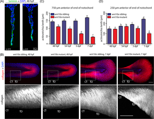
wnt10a mutants display a progressive shrinkage of MFF actinotrichia. (A) Immunofluorescence of Laminin (in green, nuclei stained with DAPI in blue), transverse sections of tail region approximately 750 μm anterior of the tip of the tail of embryos at 48 hpf. Laminin shows indistinguishable protein levels and basement membrane localization in wnt10a sibling and mutant ventral MFFs. (B) Actinotrichia, visualized via Col II immunofluorescence (in red in upper row and white in lower row; nuclear DNA stained with DAPI in blue) in the ventral MFF of wnt10a mutants and siblings at 48 hpf and 7 dpf. Lower row shows magnified views of regions framed in upper row. (C, D) Graphical illustration of lengths of actinotrichia at two corresponding locations indicated in (B) in the major lobe of the ventral MFF in wnt10a siblings and mutants at 48 hpf, 54 hpf, 3 dpf, and 7 dpf. MFF actinotrichia continue to grow in the sibling, whereas they become progressively shorter in the mutant. |