
Fig. 1
- ID
- ZDB-FIG-240619-1
- Publication
- Morooka et al., 2024 - Angpt1 binding to Tie1 regulates the signaling required for lymphatic vessel development in zebrafish
- Other Figures
- All Figure Page
- Back to All Figure Page
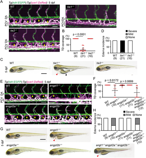
Angpt1 and Tie1 are essential for trunk lymphangiogenesis in zebrafish. (A) Representative confocal images of the trunk of Tg(kdrl:EGFP);Tg(lyve1:DsRed) WT, tie1−/− and tie2−/− larvae (5 dpf). These Tg larvae express EGFP (green) in all endothelial cells (ECs) and DsRed (magenta) in venous and lymphatic ECs. Formation of the thoracic duct (TD; yellow arrowheads in WT and tie2−/− larvae) was impaired in tie1−/− larvae. Lateral views, anterior to the left. (B) Percentage of TD coverage at 5 dpf. Data are mean±s.d. (WT, n=9 larvae; tie1−/−, n=21 larvae; tie2−/−, n=10 larvae). (C) Overall morphology of WT, tie1−/− and tie2−/− larvae (5 dpf). Red and black arrowheads point to edema around the heart and intestine, respectively. (D) Percentage of edema incidence at 5 dpf. Data are mean±s.d. (WT, n=9 larvae; tie1−/−, n=21 larvae; tie2−/−, n=10 larvae). Typical pictures of each category are shown in Fig. S2G. (E) Trunk of Tg(kdrl:EGFP);Tg(lyve1:DsRed) WT, angpt1−/−, angpt2a−/− and angpt2b−/− larvae (5 dpf). Formation of the TD (yellow arrowheads in WT, angpt2a−/− and angpt2b−/− larvae) was impaired in the angpt1−/− larvae. (F) Percentage of TD coverage at 5 dpf. Data are mean±s.d. (WT, n=9 larvae; angpt1−/−, n=10 larvae; angpt2a−/−, n=11 larvae; angpt2b−/−, n=11 larvae; angpt1−/−angpt2a−/−angpt2b−/−, n=12 larvae). (G) Overall morphology of WT, angpt1−/−, angpt2a−/−, angpt2b−/− and angpt1−/−angpt2a−/−angpt2b−/− triple-mutant larvae (5 dpf). Arrowheads point to edema around the heart. (H) Percentage of edema incidence at 5 dpf, as in D. Data are mean±s.d. (WT, n=9 larvae; angpt1−/−, n=10 larvae; angpt2a−/−, n=11 larvae; angpt2b−/−, n=11 larvae; angpt1−/−angpt2a−/−angpt2b−/−, n=12 larvae). Scale bars: 50 μm (A,E); 500 µm (C,G). P-values were determined by Kruskal–Wallis test with Dunn's test (B,F). DA, dorsal aorta; PCV, posterior cardinal vein. |