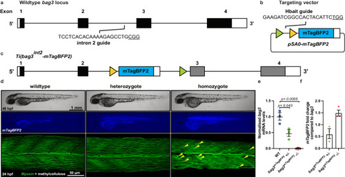
FIGURE

Fig. 2

ID
ZDB-FIG-240614-47
Publication
Miles et al., 2024 - CRIMP: a CRISPR/Cas9 insertional mutagenesis protocol and toolkit
Other Figures
All Figure Page
Back to All Figure Page
Fig. 2

Site-specific integration of targeting vector into bag3.

a Schematic of bag3 gene structure, and location of the intron 2 guideRNA site. b Schematic of pSA0-mTagBFP2 targeting vector, FRT and FRT3 sites are indicated by triangles. c Schematic of Ti(bag3int2-mTagBFP2) targeted insertion line. d Brightfield and fluorescent images of wildtype, heterozygote, and homozygote embryos at 24 hpf. Confocal images demonstrating loss of bag3 results in broken muscle fibres (yellow arrows) in homozygote mutants at 24 hpf after a 1 h 1% methylcellulose treatment. e mRNA expression analysis of bag3 demonstrates a complete loss of native bag3 transcript in homozygote mutants. f Fold change of mTagBFP2 mRNA levels in bag3mTagBFP2 heterozygous and homozygous embryos compared to bag3 levels in wildtype siblings. Error bars represent SEM for n = 3 independent biological replicates, each consisting of a pooled sample of 16 or 19 embryos. rpl13 and ef1α were used as the reference genes. Source data are provided as a Source Data file. Statistical differences were determined using a two-tailed unpaired t-test.

Expression Data

Expression Detail
Antibody Labeling
Phenotype Data

Phenotype Detail
Acknowledgments
This image is the copyrighted work of the attributed author or publisher, and ZFIN has permission only to display this image to its users. Additional permissions should be obtained from the applicable author or publisher of the image. Full text @ Nat. Commun.