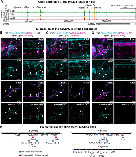
snATAC-seq identifies four lymphatic prox1a enhancers. (A) Chromatin state surrounding the prox1a locus in lymphatic endothelial cells (LECs), venous endothelial cells (VECs) and arterial endothelial cells (AECs) at 4 dpf, showing the region between 32,815,787−32,975,788 base pairs of chromosome (chr) 17. Orange, tested enhancers; purple, identified accessible chromatin sequences in LECs. (B) Confocal projections of the facial and trunk lymphatics labelled with Tg(−87prox1a:EGFP; XCA:DsRed2)uom122 (cyan) and Tg(prox1a:RFP)nim5 (magenta) at 5 dpf. Arrowheads show expression in the face and trunk lymphatics. (C) Confocal projections of the facial and trunk lymphatics labelled with Tg(−71prox1a:EGFP; XCA:DsRed2)uom121 (cyan) and Tg(prox1a:RFP)nim5 (magenta) at 5 dpf. Arrowheads show expression in the face and trunk lymphatics. Asterisks show expression in PCV. (D) Confocal projections of the facial and trunk lymphatics labelled with Tg(−14prox1a:EGFP; XCA:DsRed2)uom120 (cyan) and Tg(prox1a:RFP)nim5 (magenta) at 5 dpf. Arrowheads show expression in the facial lymphatics. (E) Predicted endothelial TF binding sites in −87prox1a. (P<1e-04), −71prox1a (P<1e-04) and −14prox1a (P<1e-04). Blue, binding sites identified in zebrafish; red, conserved binding sites within Actinopterygii. Scale bars: 50 μm.
|
