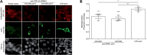
TDP-43–mediated SOD1 misfolding is attenuated by anti-RRM1 single chain antibodies.A, representative image of endogenous SOD1 misfolding (3H1 antibody, red) in HEK293 cells co-transfected with WT TDP-43 (gray) and anti-RRM1 scFvs (green) (VH1Vk9 or VH7Vk9), control scFv (D1.3, anti-chicken lysozyme), or empty vector for scFv. Scale bar represents 10 μm. B, quantification of misfolded SOD1. Data are expressed as mean ± SEM (five fields of view were quantified per condition; n = 3 experiments per condition, represented as dots in the bar graph). For each field of view, the integrated density of misfolded SOD1 (3H1) was measured and normalized to the total number of cells. Normalized misfolded SOD1 in scFv-transfected cells was then expressed as fold change compared to cells transfected with the empty vector (no scFv). Statistical significance was established using one-way ANOVA followed by Tukey’s multiple comparison test, ∗p < 0.05, ∗∗p < 0.01, and ∗∗∗p < 0.001 versus empty vector (∗) or control scFv antibody (#). RRM, RNA recognition motif; SOD1, superoxide dismutase; TDP, TAR DNA-binding protein.
|
