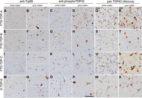
Trp68 is exposed in TDP-43 aggregates in gray and white matter in TDP43 subtypes of FTD. TDP-43 pathology in sections from the anterior cingulate cortex of FTD-TDP subtypes A, B, C, and E immunostained with anti-Trp68 (left column), commercial phospho-TDP-43 (central column), and commercial pan-TDP-43 (right column) in paraffin-embedded tissue. FTD-TDP-A shows thread-like inclusions in the white matter using the anti-Trp68 antibody (A) and the commercial pTDP-43 antibody (C). In the gray matter, cytoplasmic inclusions are predominantly stained using the anti-Trp68 antibody (B) and the pTDP-43 antibody (D) that shows abundant staining of cytoplasmic and extracellular aggregates. In FTD-TDP-B, mainly cytoplasmic inclusions are observed in the white and gray matter using the anti-Trp68 antibody (E and F) and the commercial pTDP-43 antibody (G and H). FTD-TDP-C is characterized by long threads in the white and gray matter. These long threads are seen using the anti-Trp68 antibody (I and J) and the pTDP-43 antibody (K and L). FTD-TDP-E is characterized by cytoplasmic and abundant granular TDP-43 aggregation. Using the anti-Trp68 antibody, cytoplasmic and thread-like inclusions are seen in white and gray matter (M and N), but no granular staining is observed. The pTDP-43 antibody shows cytoplasmic and granular staining in the white and gray matter (O and P). The right column (Q–X) shows pan-TDP-43 distribution, which does not well correspond to pathological antibodies. Scale bar represents 50 μm. FTD, frontotemporal domain; TDP, TAR DNA-binding protein.
|
