FIGURE

FIGURE 4

ID
ZDB-FIG-240511-10
Publication
Sneider et al., 2024 - Small extracellular vesicles promote stiffness-mediated metastasis
Other Figures
All Figure Page
Back to All Figure Page
FIGURE 4

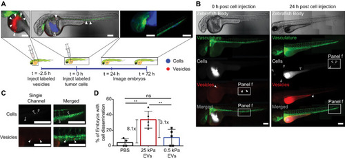
Stiff EVs promote cancer cell migration and dissemination. A, Schematic of 2-day GFP+ zebrafish embryo model system used to test the ability of MDA-MB-231 EVs (red, filled triangles) to disperse MDA-MB-231 cancer cells (blue, open triangles). Scale bar, 100 µm. B, Full zebrafish body images of the embryos at time 0 and 24 hours postinjection of MDA-MB-231 cancer cells. Scale bar, 100 µm. Representative images. C, Enlarged images of disseminated MDA-MB-231 cells (white) and EVs (red). Scale bar, 100 µm. Representative images. D, Percentage of injected embryos with cancer cell dissemination to the head or the tail. Total number of fish per condition is 47 for PBS control, 63 for 25 kPa, and 78 for 0.5 kPa condition. Five biological repeats of EVs. One-way ANOVA.

Expression Data

Expression Detail
Antibody Labeling
Phenotype Data

Phenotype Detail
Acknowledgments
This image is the copyrighted work of the attributed author or publisher, and ZFIN has permission only to display this image to its users. Additional permissions should be obtained from the applicable author or publisher of the image. Full text @ Cancer Res Commun