Fig. 3
- ID
- ZDB-FIG-240509-80
- Publication
- DeSmidt et al., 2019 - Zebrafish Model for Non-Syndromic X-linked Sensorineural Deafness, DFNX1
- Other Figures
- All Figure Page
- Back to All Figure Page
|
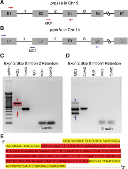
Efficacy of antisense morpholino knockdown of prps1a and prps1b in zebrafish. (A) Schematic drawing of genomic structure of the prps1a gene in chromosome 5 with PCR primers (red arrows) and morpholino 1 (MO1, red bar) targeting the junction between exon 2 (E2) and intron 2 (I2). (B) Schematic drawing of genomic structure of the prps1b gene in chromosome 14 with PCR primers (blue arrows) and morpholino 2 (MO2, blue bar) targeting the junction between intron 1 and exon 2. (C) DNA gel of RT-PCR products for MO1 morphants shows multiple bands between the red arrowhead and the red arrow compared with one band for control MO (CoMO) zebrafish. (D) DNA gel of RT-PCR products for MO2 morphants also shows multiple bands between the blue arrowhead and the blue arrow compared with one band for CoMO zebrafish. H2O: negative control, and ß-actin: positive control. (E) A portion of the sequence of the DNA gel enclosed by the red box in C for MO1 morphants, showing the full length of I2 retention (highlighted in red) for MO1 morphants. Sixty DNA bases of E2 upstream I2 and 60 bases of E3 downstream I2 of prps1a are highlighted in yellow. |

