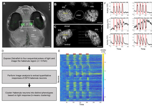
The habenula has multiple subtypes of cells that show differential response to light. (A) Dorsal view of the head of a live 7-day-old fish, with GCaMP3 expression in the habenula (arrows) under the control of the s1011t GAL4 driver. (B) A single two-photon slice through the dorsal habenula of the fish in panel A (boxed region). (C) A yz-reconstruction at the point indicated by the yellow line in panel B, showing a transverse view of the habenula. The dotted lines indicate imaging planes separated by 10 μm. The yellow line indicates the plane imaged in B. Dashed lines show the border of the habenula. (D) Workflow of habenula analysis (E) Heat map showing the activity of 2974 individual neurons (rows) across multiple light on and off cycles. Red bars on top correspond to the time period when light is switched on. As seen, some neurons show higher activity during light exposure (ON cells) and some neurons show higher activity after the light is switched off (OFF cells). (F) Response (y-axis) vs. time (x-axis) for habenula neurons that were clustered into subgroups depending on their response to light. Pink regions in each plot corresponds to the light exposure period. Seen here are the three subtypes in the dorsal habenula region (left) and three in the ventral habenula region (right). In total six subtypes were identified: D-ON-Tonic, D-OFF-Tonic, D-OFF-Phasic, V-ON-Tonic, V-OFF-Tonic, and V-OFF-Phasic. lHb: left habenula; rHb: right habenula; a: anterior; p: posterior. Scale bar = 25 μm
|
