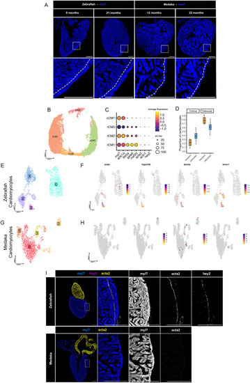
Medaka lack primordial myocardium and have few cortical layer cardiomyocytes. (A) RNA in situ hybridization of myl7 labeling myocardium in zebrafish and medaka ventricles of the indicated age. Dotted line indicates border between trabecular and cortical layers. Images are representative of at least three individuals at each time point. Scale bars: 200 µM. (B) UMAP embedding of re-clustered cardiomyocytes identified as either trabecular (tCM) or cortical (cCM). (C) Gene expression dot plot showing expression of marker genes for each CM cell cluster. (D) Proportion of cardiomyocytes in trabecular or cortical cell clusters from all single-cell samples from zebrafish and medaka. (E,F) UMAP embedding of ventricular cardiomyocytes clustered separately from uninjured (E) zebrafish or (F) medaka. (G,H) Gene expression feature plots for top marker genes for primordial cardiomyocytes in zebrafish (E) or medaka (F). (I) RNA in situ hybridization of myl7, acta2, and hey2 in uninjured zebrafish hearts. Scale bars: 200 µM. (J) RNA in situ hybridization of myl7 and acta2 in uninjured medaka heart. Scale bars: 200 µM.
|