FIGURE

Fig. 4

ID
ZDB-FIG-240328-75
Publication
Sebastian et al., 2024 - Cardiac manifestations of human ACTA2 variants recapitulated in a zebrafish model
Other Figures
All Figure Page
Back to All Figure Page
Fig. 4

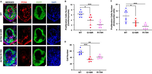
Proliferating cells were reduced in the ACTA2 G148R variant. AD Immunostaining of Tg[cmlc2:EGFP] ACTA2 larvae heart at 4 days post-fertilization (green) with proliferation cell marker (PCNA, red), 4’,6-diamidino-2-phenylindole (DAPI) co-staining (blue). Scale bar: 20 μm. A Endocardial cells (yellow arrowheads) and cardiomyocytes (white arrowheads). Zebrafish with ACTA2 p.G148R and ACTA2 p.R179H show reduced total cell numbers. B In the myocardium, ACTA2 p.G148R and ACTA2 p.R179H cardiomyocyte showed decreased numbers of proliferating cells in the heart wall area highlighted with EGFP green fluorescence, PCNA, and DAPI. C In the endocardium area, endocardium cells showed decreased proliferation in all pathogenic variants highlighted with PCNA and DAPI. **P < 0.01, ***P < 0.001. Error bars indicate the standard deviation

Expression Data

Expression Detail
Antibody Labeling
Phenotype Data

Phenotype Detail
Acknowledgments
This image is the copyrighted work of the attributed author or publisher, and ZFIN has permission only to display this image to its users. Additional permissions should be obtained from the applicable author or publisher of the image. Full text @ J. Hum. Genet.