FIGURE SUMMARY
Title

Cardiac manifestations of human ACTA2 variants recapitulated in a zebrafish model

Authors
Sebastian, W.A., Inoue, M., Shimizu, N., Sato, R., Oguri, S., Itonaga, T., Kishimoto, S., Shiraishi, H., Hanada, T., Ihara, K.
Source
Full text @ J. Hum. Genet.

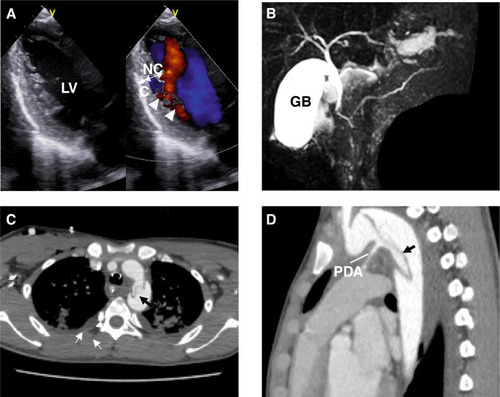
Echocardiography and magnetic resonance cholangio-pancreatography (MCRP) of the patient with the ACTA2 G148R variant. A An echocardiogram showing the left ventricular long-axis view during diastole. The ventricular wall of the left ventricle exhibits an outer compacted layer (C) and an inner non-compacted trabecular layer (NC), with deep recesses formed by the trabeculations (NC/C ratio 2.0). Color Doppler imaging shows blood infiltrating into the spaces between the trabeculae (arrowheads). B On MRCP, the main pancreatic duct exhibits a reverse Z-shape (arrows) in the head of the pancreas region. C, D Chest magnetic resonance imaging shows the descending aortic deviation. LV left ventricle, C compacted layer, NC non-compacted layer, GB gallbladder, PDA patency of the ductus arteriosus

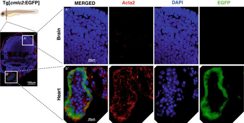
Endogenous ACTA2 expression in the heart. Schematic figure of transversal section of zebrafish larvae Tg[cmlc2:EGFP] at 4 days post-fertilization. Immunostaining of 4dpf larvae Tg[cmlc2:EGFP] with anti-acta2 antibody (red), followed by 4’,6-diamidino-2-phenylindole (DAPI) co-staining (blue). A’ Brain region. B’ Heart region. Scale bar: 100 μm. A”, B” High magnification of immunostaining image. Scale bar: 20 μm. A” In the brain area, endogenous ACTA2 was not identified. B” In the heart area, endogenous ACTA2 was highly expressed, highlighted with EGFP green fluorescence, anti-acta2 antibody (red), and DAPI. Illustration of zebrafish was created with Biorender.com

EXPRESSION / LABELING:
Gene:
Antibody:
Fish:
Anatomical Term:
Stage: Day 4

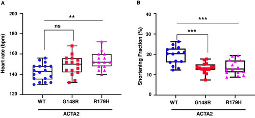
Heart contraction impairs in the ACTA2 G148R variant. A, B Heart rate and shortening fraction measurement at 3 days post-fertilization. A Zebrafish with ACTA2 p.G148R and ACTA2 p.R179H show an increased heart rate (beats per minute). B Shortening fractions were significantly reduced in ACTA2 G148R and ACTA2 R179H. *P < 0.05, ***P < 0.001

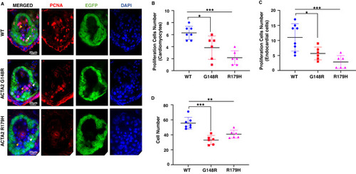
Proliferating cells were reduced in the ACTA2 G148R variant. AD Immunostaining of Tg[cmlc2:EGFP] ACTA2 larvae heart at 4 days post-fertilization (green) with proliferation cell marker (PCNA, red), 4’,6-diamidino-2-phenylindole (DAPI) co-staining (blue). Scale bar: 20 μm. A Endocardial cells (yellow arrowheads) and cardiomyocytes (white arrowheads). Zebrafish with ACTA2 p.G148R and ACTA2 p.R179H show reduced total cell numbers. B In the myocardium, ACTA2 p.G148R and ACTA2 p.R179H cardiomyocyte showed decreased numbers of proliferating cells in the heart wall area highlighted with EGFP green fluorescence, PCNA, and DAPI. C In the endocardium area, endocardium cells showed decreased proliferation in all pathogenic variants highlighted with PCNA and DAPI. **P < 0.01, ***P < 0.001. Error bars indicate the standard deviation

Acknowledgments
This image is the copyrighted work of the attributed author or publisher, and ZFIN has permission only to display this image to its users. Additional permissions should be obtained from the applicable author or publisher of the image. Full text @ J. Hum. Genet.