FIGURE

Fig. 2.

ID
ZDB-FIG-240319-47
Publication
Jackman et al., 2024 - Blocking endogenous retinoic acid degradation induces oral tooth formation in zebrafish
Other Figures
All Figure Page
Back to All Figure Page
Fig. 2.

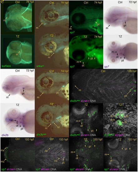
TZ exposure changes the position of the mouth and induces oral tooth formation in zebrafish. TZ treatment starting at 24 hpf results in a posteriorly positioned mouth by 78 hpf (surface staining A and B; shhKI reporter C and D). Tooth germs are seen surrounding the mouth as visualized by the sp7 dental mesenchyme marker (GFP reporter E and F; mRNA ISH G and H) and dental epithelium marker dlx2b (mRNA ISH I and J; GFP reporter K and L). By 128 hpf, compared with a control lacking oral tooth germs (M), a TZ-exposed larva exhibits oral dental epithelial reporter expression as well as partial mineralization seen with alizarin red S staining (N and N’). By 150 hpf, compared with a control lacking oral tooth germs (O and P), a TZ-exposed larva exhibits oral dental epithelial reporter expression in a more anteriorly positioned mouth (Q and R). However, at this stage, we detect no evidence of partial mineralization. Boxed region in N is zoomed in for N’. Abbreviations: cb: ceratobranchial, m = mouth, mc = Meckel’s cartilage, mx = maxilla, op = opercle, ot = oral tooth germs/teeth, pt = pharyngeal tooth germs/teeth.

Expression Data

Expression Detail
Antibody Labeling
Phenotype Data

Phenotype Detail
Acknowledgments
This image is the copyrighted work of the attributed author or publisher, and ZFIN has permission only to display this image to its users. Additional permissions should be obtained from the applicable author or publisher of the image. Full text @ Proc. Natl. Acad. Sci. USA