
FIGURE
Fig. 1.
- ID
- ZDB-FIG-240319-46
- Publication
- Jackman et al., 2024 - Blocking endogenous retinoic acid degradation induces oral tooth formation in zebrafish
- Other Figures
- All Figure Page
- Back to All Figure Page
Fig. 1.
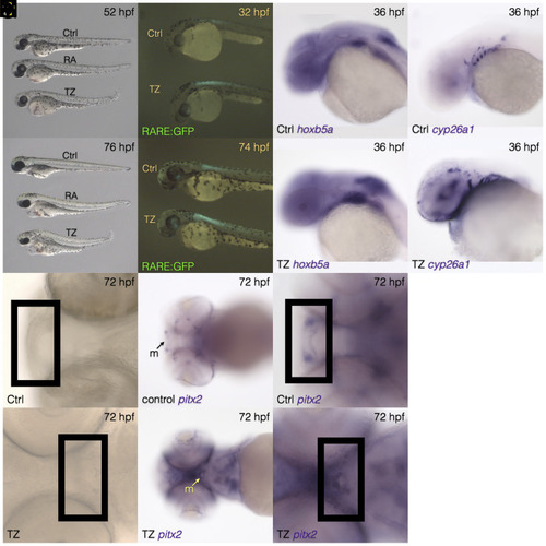
Morphological and gene expression changes induced by TZ exposure. Whole body morphology is similar between embryos exposed to exogenous RA and to TZ starting at 24 hpf ( |
Expression Data
Expression Detail
Antibody Labeling
Phenotype Data
Phenotype Detail
Acknowledgments
This image is the copyrighted work of the attributed author or publisher, and
ZFIN has permission only to display this image to its users.
Additional permissions should be obtained from the applicable author or publisher of the image.
Full text @ Proc. Natl. Acad. Sci. USA