FIGURE

Figure 2

ID
ZDB-FIG-240215-98
Publication
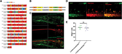
Raas et al., 2024 - Generation and characterization of a zebrafish gain-of-function ACOX1 Mitchell disease model
Other Figures
All Figure Page
Back to All Figure Page
Figure 2

(A) Zebrafish Acox1 protein is highly conserved compared to human ACOX1. The degree of amino acid similarity is indicated by color, with red being identical and blue being non-conserved, as visualized using PRALINE. (B) Schematic diagram of expression constructs used in zebrafish experiments. (C) Confocal z-stack images of the expression of the construct in live zebrafish larvae; immunohistochemistry for anti-GFP, and anti-RFP in the Tg(olig2:dsRed) line. Dorsal to top, rostral to the right; age 3 dpf. Note: the black line in the middle of the figure indicates where the image was stitched together. (D) Confocal z-stack images, spinal cord, rostral to the right, dorsal to the top. Expression of N237S is indicated by tagged GFP, and mature oligodendrocytes are labeled by olig2:dsRed. (E) Quantification shows no effect on oligodendrocyte numbers.

Expression Data

Expression Detail
Antibody Labeling
Phenotype Data

Phenotype Detail
Acknowledgments
This image is the copyrighted work of the attributed author or publisher, and ZFIN has permission only to display this image to its users. Additional permissions should be obtained from the applicable author or publisher of the image. Full text @ Front Pediatr