
Fig. 2
- ID
- ZDB-FIG-240215-124
- Publication
- Peles et al., 2022 - Glucocorticoid-sensitive period of corticotroph development-Implications for mechanisms of early life stress
- Other Figures
- All Figure Page
- Back to All Figure Page
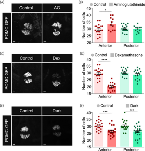
Perturbations of cortisol levels cause alterations in proopiomelanocortin (POMC) cell clusters. (A, B) Larvae were treated with 0.2 mM aminoglutethimide (AG), a cortisol synthesis inhibitor, from 3 to 5 days post fertilization (dpf). Treated and untreated control larvae were collected at 6 dpf and fixed. The pituitary was imaged (A) and the intensity of POMC-positive cells in the anterior and posterior clusters was analyzed. (B) Unpaired t-test comparing anterior and posterior clusters independently, n = 19 control/11 AG– treated. (C, D) Larvae were treated with 35 μM dexamethasone (Dex), a cortisol analogue, from 1 to 5 dpf. Treated and untreated control larvae were collected at 6 dpf and fixed. The pituitary was imaged (C) and the intensity of POMC-positive cells in the anterior and posterior clusters was analyzed. (D) Unpaired t-test comparing anterior and posterior clusters independently, n = 15 per group). (E, F) Larvae were grown in the dark from 1 to 5 dpf. Treated and control larvae (grown in the presence of light–dark cycles) were collected and fixed. The pituitary was imaged (E) and the intensity of POMC-positive cells in the anterior and posterior clusters was analyzed. (F) Unpaired t-test comparing anterior and posterior clusters independently, n = 21 per group. Data presented as mean ± SEM, *p < .05, ***p < .001, ****p < .0001, scale bars: 10 μm. |