
Fig. 4
- ID
- ZDB-FIG-240208-9
- Publication
- Saul et al., 2023 - Optogenetic Signaling Activation in Zebrafish Embryos
- Other Figures
- All Figure Page
- Back to All Figure Page
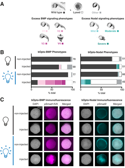
Assessing light-activated signaling responses in zebrafish embryos. Zebrafish embryos were injected at the one-cell stage with mRNA encoding bOpto-BMP/Nodal. (A) Embryos were either reared in the dark or exposed to uniform blue light starting at 1.5 h post fertilization (hpf). Phenotypes were scored at 1 day post fertilization (dpf). Representative phenotypes are shown. Excess BMP signaling leads to ventralization (left panel), while excess Nodal signaling causes developmental defects associated with extra mesendoderm (right panel). Scale bar = 500 µm. (B) Phenotype quantification. Injected embryos and non-injected siblings were reared in the dark starting at 1.5 hpf (black bulb). Half of the injected and half of the non-injected embryos were exposed to uniform blue light (blue bulb). (C) Injected embryos and non-injected siblings were reared in the dark starting at 1.5 hpf (black bulb). At 40% epiboly (~6 hpf), half of the injected and half of the non-injected embryos were exposed to uniform blue light (blue bulb). After 20 min, all embryos were fixed and subjected to immunofluorescence staining for either phosphorylated Smad1/5/9 or Smad2/3. Higher pSmad intensities indicate increased BMP/Nodal signaling, respectively. Scale bar = 200 µm. |