
Fig. 3
- ID
- ZDB-FIG-240208-8
- Publication
- Saul et al., 2023 - Optogenetic Signaling Activation in Zebrafish Embryos
- Other Figures
- All Figure Page
- Back to All Figure Page
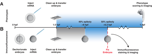
bOpto-BMP/Nodal experiment workflow. Phenotype assay and pSmad immunofluorescence staining to test activity of bOpto-BMP/Nodal. Embryos are injected with mRNA at the one-cell stage and transferred to a light box no later than 1.5 h post-fertilization (hpf). (A) Phenotype assay. Injected embryos and non-injected siblings are reared in the dark or exposed to uniform blue light starting at 1.5 hpf until 1-day post-fertilization (dpf). Optogenetic signaling activity can be evaluated by scoring embryos for phenotypes consistent with excess pathway activity. (B) pSmad immunofluorescence staining. Injected embryos and non-injected siblings are reared in the dark until 40% epiboly (~6 hpf). Half of the injected and half of the non-injected embryos are then exposed to uniform blue light for 20 min. After exposure, all embryos are fixed and subjected to immunofluorescence staining for pSmad. Elevated levels of pSmad1/5/9 or pSmad2/3 reflect optogenetic activation of BMP or Nodal signaling, respectively. |