
FIGURE
Fig. 4
- ID
- ZDB-FIG-240202-35
- Publication
- Nurcombe et al., 2023 - Plexina4 and cell survival in the developing zebrafish hindbrain
- Other Figures
- All Figure Page
- Back to All Figure Page
Fig. 4
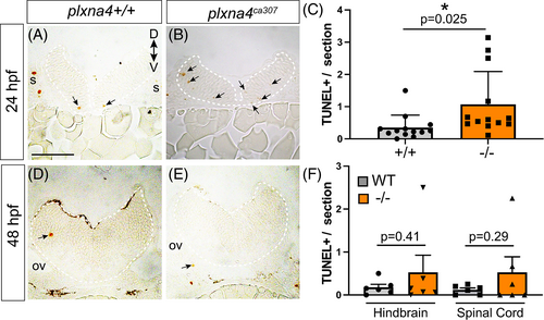
Apoptotic cell death in the plxna4ca307 hindbrain. (A–D) Representative hindbrain sections show TUNEL+ dying cells (arrows) in 24 (A, B) and 48 (D, E) hpf WT and mutant embryos. (C, F) Blinded quantitation of average number of TUNEL+ cells/section in the hindbrain of WT and plxna4ca307 embryos at 24 hpf (N = 5; WT n = 13; −/− n = 13; unpaired Student's t-test, P = 0.0251) and 48 hpf hindbrain and spinal cord (N = 2; WT n = 6, plxna4ca307 n = 5, unpaired Student's t-test HB, P = 0.4061. SC, P = 0.2889. D: dorsal, ov: otic vesicle, V: ventral. Scale bar in A is 75 μm. |
Expression Data
Expression Detail
Antibody Labeling
Phenotype Data
| Fish: | |
|---|---|
| Observed In: | |
| Stage: | Prim-5 |
Phenotype Detail
Acknowledgments
This image is the copyrighted work of the attributed author or publisher, and
ZFIN has permission only to display this image to its users.
Additional permissions should be obtained from the applicable author or publisher of the image.
Full text @ Dev. Dyn.