
Fig. 6
- ID
- ZDB-FIG-240130-6
- Publication
- Hu et al., 2023 - Intraocular Axon Regeneration in a Model of Penetrating Eye Injury
- Other Figures
- All Figure Page
- Back to All Figure Page
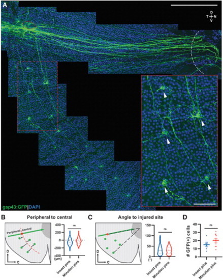
Ventral RGCs project axons circumferentially to the injury site. (A) Representative retina flat-mount image from a 3-day postinjury retina with magnification showing GFP+ RGCs (white arrowheads) from the ventral side of the injury projecting axons to the injury site (red circle: injured site, white curve: optic nerve head). (B) Schematic description and quantification of circumferential projecting GFP+ RGCs distribution relative to the injury site. (C) Schematic description and quantification of circumferential GFP+ RGCs relative angle to injury site. (D) Mean number of circumferential GFP+ cells activated postretina penetrating injury. In (B, C), 126 GFP+ cells from insect pins and 215 GFP+ cells from Minutien pins group were measured for violin plot. Data in (D) are presented as mean ± SEM. All data were acquired from ∼5 retinas, 3 animals. ns by Student's t-test. Scale bar = 200 and 50 μm. |