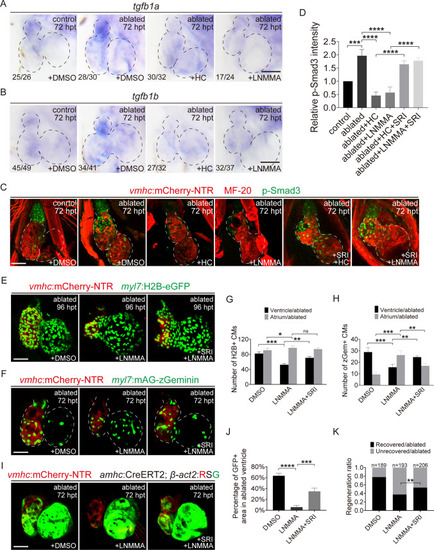
NO signaling mediates ventricle regeneration through TGF-β pathway. A, B Whole-mount in situ hybridizations showed the expression of tgfb1a and tgfb1b at 72 hpt in control and ablated hearts treated with DMSO, HC-067047 or L-NMMA for 48–72 hpt. Numbers indicate the ratio of representative staining observed. C Immunostaining of anti-myosin heavy chain (MF-20, red) and anti-phospho-Smad3 (green) in control and ablated Tg(vmhc:mCherry-NTR) hearts treated with DMSO, HC-067047 or L-NMMA alone or with SRI-011381 for 48–72 hpt. D Quantification of relative p-Smad3 intensity in control or ablated hearts with DMSO, HC-067047, L-NMMA or SRI-011381 treatment for 48–72 hpt. N = 7 for each group. Mean + s.e.m. ANOVA analysis; ***P < 0.001, ****P < 0.0001. E, F Confocal images of ablated hearts treated with DMSO, L-NMMA or L-NMMA + SRI-011381 for 48–72 hpt in Tg(vmhc:mCherry-NTR; myl7:H2B-eGFP) larvae at 96 hpt and in Tg(vmhc:mCherry-NTR; myl7:mAG-zGeminin) larvae at 72 hpt. G, H Quantification of H2B+ CM and zGeminin+ CM numbers in DMSO-, L-NMMA- and L-NMMA + SRI-011381-treated ablated hearts. N = 10 for each group. Mean + s.e.m. ANOVA analysis; ns, not significant; *P < 0.05, **P < 0.01, ***P < 0.001. I Confocal images of ablated hearts treated with DMSO, L-NMMA or L-NMMA + SRI-011381 for 48–72 hpt in Tg(vmhc:mCherry-NTR; amhc:CreERT2; β-act2:RSG) larvae at 72 hpt. J Quantification of the percentages of GFP-positive area in DMSO-, L-NMMA- and L-NMMA + SRI-011381-treated ablated hearts. N = 10 for each group. Mean + s.e.m. ANOVA analysis; ***P < 0.001, ****P < 0.0001. K Quantification of the heart recovery rate in ablated groups treated with DMSO, L-NMMA, L-NMMA + SRI-011381 at 96 hpt. The numbers of larvae analyzed for each condition are indicated. Binomial test, **P < 0.01. Scale bars, 50 μm. Dashed lines outline the hearts. hpt hours post treatment, CM cardiomyocyte, HC HC-067047, SRI SRI-011381
|
