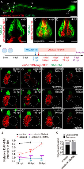
Inhibition of nitric oxide synthesis impedes zebrafish ventricle regeneration. A DAF-FM DA staining showed that NO (green) was primarily present in the bulbus arteriosus (BA), cleithrum (C), pharyngeal jaw bone (P), notochord (N) and caudal fin (CF) in larvae at 4 dpf. Scale bar, 150 μm. B–D DAF-FM DA staining of Tg(vmhc:mCherry-NTR) or Tg(kdrl:mCherry-ras) hearts at 4 dpf showed that NO was enriched in the smooth muscle layer of the BA. Scale bars, 10 μm. E Schematic timeline diagram of MTZ treatment to induce ventricle ablation and L-NMMA treatment to inhibit NO production. F–I″ The NO level was significantly decreased in ablated hearts at 24 hpt and gradually increased to a level comparable to control hearts at 96 hpt (F–G″). L-NMMA treatment dramatically reduced NO level in control and ablated hearts (H–I″). Dashed lines outline the hearts. Scale bars, 50 μm. J Quantification of relative DAF-FM DA intensity of BA in control or ablated hearts with or without L-NMMA treatment. N = 5 for each group. Mean + s.e.m. ANOVA analysis, ***P < 0.001, ****P < 0.0001 as compared with control group; ##P < 0.01, ###P < 0.001, ####P < 0.0001 as compared with ablated group. K Quantification of the heart recovery rate in the ablated and ablated + L-NMMA-treated groups at 96 hpt. The numbers of larvae analyzed for each condition are indicated. Binomial test, ***P < 0.001, ****P < 0.0001. dpf days post fertilization, hpt hours post treatment, V ventricle, BA bulbus arteriosus, NO nitric oxide
|
