|
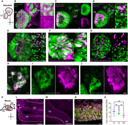
foxj1 is expressed in ciliated OSNs of zebrafish and mice. (A) Schematic showing the larval zebrafish OE and OB; l: lateral, m: medial, d: dorsal, v: ventral. (B-J) Confocal images of the larval (3–5 dpf) zebrafish nose and OB. (B) foxj1b (Gt(foxj1b:GFP), green) is expressed in neurons labeled by HuC (magenta). Asterisks show foxj1-expressing neurons in 5 dpf larva. (C) Differential expression of foxj1a (Gt(foxj1a:2A-TagRFP), magenta) and foxj1b (Gt(foxj1b:GFP), green) paralogs in 5 dpf larvae. Note that foxj1a is expressed primarily at the outer rim of the OE. (D) foxj1b-positive cells (Gt(foxj1b:GFP), green) bear a brush of motile cilia marked by glutamylated-tubulin (magenta) in 5 dpf larvae. (E) foxj1b-positive OSNs (Gt(foxj1b:GFP), green) in the nasal pit bear cilia, indicated by the marker Gαolf (magenta) in 3 dpf larva. White arrowheads in insets show Gαolf marked cilia arising from foxj1b-positive OSNs. (F) Overlap of foxj1b-expressing cells (Gt(foxj1b:GFP), green) with the ciliated OSN marker OMP (Tg(OMP:gal4); Tg(UAS:NTR-mCherry), magenta) in 4 dpf larva. (G) Mutually exclusive localization of foxj1b-expressing cells (Gt(foxj1b:GFP), green) and microvilli OSNs in 4 dpf larvae, indicated by the marker trpc2 (Tg(trpc2:gal4); Tg(UAS:NTR-mCherry), magenta). (H) foxj1b-expressing OSNs (Gt(foxj1b:GFP), green) project mainly to Gαolf-labeled glomeruli (magenta) in 3 dpf larvae. Note that foxj1b:GFP and Gαolf do not overlap entirely (dotted region). (I, J) foxj1b-positive OSNs, in 4 dpf larvae, (I) project to more glomeruli than OMP-expressing OSNs (J) (Tg(OMP:ChR-YFP)). Glomeruli are shown by staining with the presynaptic vesicle marker SV2 (magenta). (K) Schematic showing mouse RE, OE, and OB; r: rostral, c: caudal, d: dorsal, v: ventral. (L) In the nasal epithelium of the adult mouse (P30), Foxj1 localizes to MCCs (arrowhead) within the RE and the layer of OSNs in the OE. Dashed line demarcates the lamina propria separating OE from the underlying tissue. (M) Foxj1 is absent from the nasal epithelium of the Foxj1 knockout mouse (P21). The residual puncta likely represent nonspecific staining of blood vessels or mesenchymal tissue located in the lamina propria under the basement membrane. (N) Foxj1 (magenta) localizes to the nuclei of mature OSNs immunostained for the OMP (P30). (O) Expression levels of Foxj1 in OSNs at different ages (newborn P0, day 5 P5, and adult P30) calculated by comparing immunofluorescence in OSNs relative to respiratory MCCs. No significant difference was found (MCC/OSN ratio of 10.87 ± 2.61, P0; 8.74 ± 0.53, P5; 6.83 ± 1.56, P30; fluorescence intensity was measured in 20–30 cells of each type per individual field of view and ratio was calculated per animal, 3 mice in each group) by one-way ANOVA with Kruskal–Wallis test. Scale bars = 10 μm (B-J), 50 μm (L, M), and 10 μm (N). Raw data files are available in Mendeley Data (https://data.mendeley.com/datasets/2pn963jn6y). dpf, days post fertilization; MCC, motile multiciliated cell; OB, olfactory bulb; OE, olfactory epithelium; OMP, olfactory marker protein; OSN, olfactory sensory neuron; RE, respiratory epithelium.
|