
Fig. 1
- ID
- ZDB-FIG-240112-73
- Publication
- Travisano et al., 2023 - Single-nuclei multiomic analyses identify human cardiac lymphatic endothelial cells associated with coronary arteries in the epicardium
- Other Figures
- All Figure Page
- Back to All Figure Page
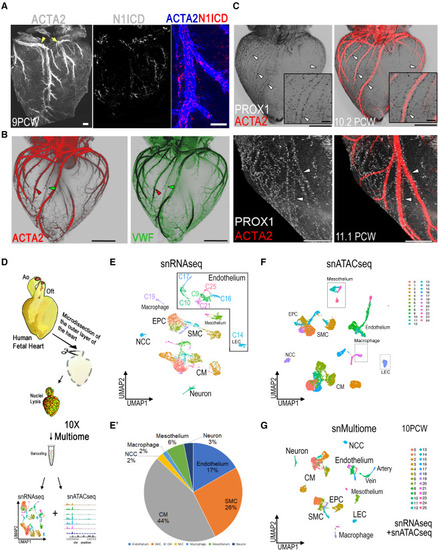
snMultiomic analysis of human cardiac lymphatic vessels that are mainly associated with coronary arteries (A) Whole-mount dorsal view of 9PCW human fetal hearts immunostained for ACTA2 and N1ICD. The RCA (arrowhead) and the CXA (arrow) are marked (n = 3). (B and C) Whole-mount light sheet images of the 10.2PCW and 11PCW (C, bottom) dorsal views of human fetal hearts stained for smooth muscle marker ACTA2 (red) and endothelial cell marker VWF (green) (B) and LEC-expressing PROX1 (gray) (C). Magnified views (inset) show details of the RCA. Red arrowheads in (B): CA; green arrowheads in (B): CV. White arrowheads in (C) indicate the PROX1+ cell nuclei marking LECs associated with the ACTA2+ arteries on the ventricle (n = 15). PROX1+ nuclei (lymphatic) primarily align along the artery but also, rarely, with the vein. (D) Schematic of the workflow for isolation and enrichment of epicardium before the cell dissociation to perform the 10× snMultiome profiling. Ao, aorta; OFT, outflow tract. (E) UMAP of snRNA-seq data for all cells of the 10PCW heart showing several endothelial clusters. (E′) Pie chart showing percentage of all cell clusters from the snMultiome analyses. (F) UMAP of chromatin accessibility labeled as snATAC-seq. (G) UMAP of gene expression and chromatin accessibility computed using the WNN and labeled as snMultiome. Scale bars, 200 μm. |