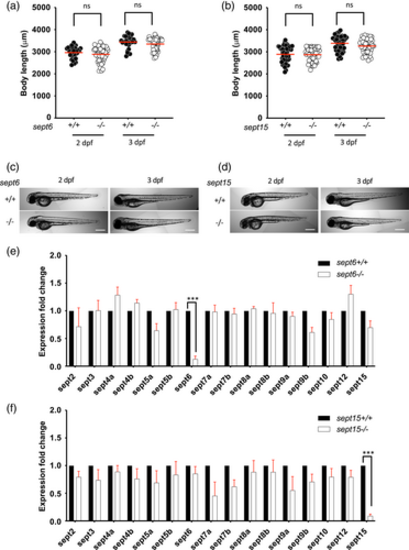
Characterisation of septin mutants. (a–d) sept6 (a,c) and sept15 (b,d) mutants do not have aberrant development. Comparison of the larvae length for sept6 (a) and sept15 (b) mutants to wild-type larvae, at 2 and 3 dpf. The body lengths of mutants and wild-types did not differ significantly. Representative images of sept6 (c) and sept15 (d) mutant and wild-type larvae at 2 and 3 dpf. Scale bar: 500 μm. Results are cumulative of three independent experiments. Sample size: in total, 30 (wild-type) and 48 (mutant) fish larvae were analysed for sept6 and 48 (wild-type) and 48 (mutant) fish larvae were analysed for sept15. Statistics: Kruskal–Wallis test; ns p > 0.05. (e,f) Both sept6 (e) and sept15 (f) homozygote mutations lead to nonsense-mediated mRNA decay of the mutated transcript but do not significantly impact the expression level of other septins. Expression analysis was performed by qRT-PCR, using primers able to amplify both mutant and wild-type transcripts. Data are represented as expression fold changes against the expression level of the wild-type group. Sample size: 15–20 larvae at 3 dpf were pooled together to extract mRNA from four independent replicates. Statistics: differences in gene expression levels were determined by multiple paired t tests, with Benjamini, Krieger, and Yekutieli to correct for false discovery (false discovery rate assumed 1%). ***p < 0.001
|

