
Fig. 5
- ID
- ZDB-FIG-231215-5
- Publication
- Tu et al., 2022 - Dhx38 regulates the maintenance and differentiation of erythro-myeloid progenitors and hematopoietic stem cells by alternative splicing
- Other Figures
- All Figure Page
- Back to All Figure Page
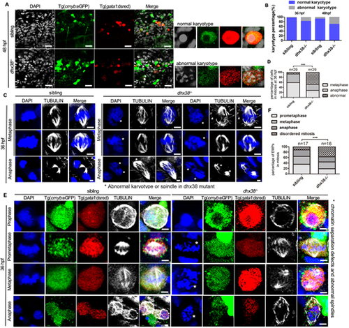
EMPs in dhx38 mutants exhibit disordered mitosis. (A) Confocal images of immunostaining of Tg(cmyb:eGFP;gata1:dsred) fish and DAPI (white) show that an abnormal karyotype occurs in the dhx38 mutants at 48 hpf. The gray boxes indicate cells with abnormal karyotypes. (B) Quantification of cells with abnormal karyotypes. For 36 hpf, number of cells with abnormal karyotypes: sibling, 32/768; dhx38−/−, 128/820. For 48 hpf, number of cells with abnormal karyotypes: sibling, 64/736; dhx38−/−, 248/552. (C) Confocal images of immunostaining for α-tubulin and DAPI. The first three panels in the wild-type siblings show a normal karyotype and spindles during metaphase and anaphase. The last six panels in dhx38−/− show a ‘grape’ karyotype in metaphase. The ‘grape’ karyotype appears to represent chromosomes unable to align at the equatorial plate, and these cells progress to disordered anaphase in dhx38−/− embryos. Asterisks indicate abnormal karyotypes or spindles. (D) Quantification of double-positive fluorescent cell number from C. (E) Confocal images of immunostaining for cmyb (green), gata1 (red), DAPI (blue) and α-tubulin (white) at 36 hpf. EMPs (cmyb+/gata1+) undergo normal prometaphase, metaphase and anaphase in siblings, but exhibit an abnormal chromatin karyotype accompanied by a progressively abnormal spindle morphology in the dhx38 mutants. Asterisks represent abnormal mitotic processes. (F) Quantification of the percentage of EMPs in mitosis from E. Scale bars: 50 μm (A); 5 μm (C,E). ***P<0.001. |