
Fig. 1
- ID
- ZDB-FIG-231215-1
- Publication
- Tu et al., 2022 - Dhx38 regulates the maintenance and differentiation of erythro-myeloid progenitors and hematopoietic stem cells by alternative splicing
- Other Figures
- All Figure Page
- Back to All Figure Page
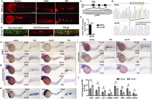
Knockout of dhx38 in zebrafish impaired EMP differentiation. (A) In vivo imaging of the expression of the CMV promoter vector and dhx38 promoter vector in wild-type embryos. Scale bars: 200 μm. The dhx38 promotor mainly drives mCherry expression in the PBI region (indicated by white arrows). (B) Representative images of dhx38 expression in Tg(cmyb:eGFP) embryos at 36 hpf. White arrows indicate the co-expression of dhx38 and cmyb. Scale bars: 50 μm. (C) A schematic diagram of the dhx38 gRNA locus. (D) DNA sequencing identified a 10 bp deletion of cDNA (c.11_21delTTCCTCCCTG), which predicts a truncated protein (p.Ser5_ThrfsTer7). (E) qRT-PCR shows a significant decrease of dhx38 mRNA in the dhx38 mutants. (F,G) WISH results show that the expression of the EMP markers lmo2 and gata1 and the promyelocyte markers pu.1 and cebpa in the dhx38−/− embryo is increased at 33 hpf. The red line denotes increased expression. Scale bars: 200 μm. (H,I) WISH results showing that the expression of the myelocyte markers mpx, mfap4 and mpeg1, and the erythrocyte marker hbae1 is decreased in the dhx38 mutant at 33 hpf. The blue lines denote decreased expression. (J) Quantification of cell number in the PBI shown in F-I. Data show the mean±s.d. Significance was determined using a two-tailed, unpaired Student's t-test. *P<0.05, **P<0.01; ****P<0.0001. |