
Fig. 1
- ID
- ZDB-FIG-231215-49
- Publication
- Lu et al., 2019 - Mcidas mutant mice reveal a two-step process for the specification and differentiation of multiciliated cells in mammals
- Other Figures
- All Figure Page
- Back to All Figure Page
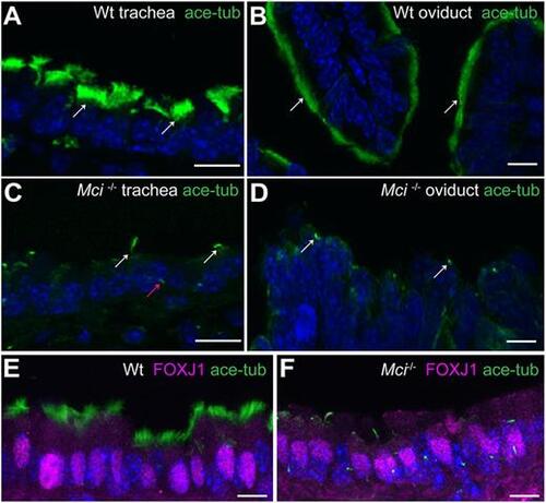
MCCs in Mci mutant mice differentiate a single cilium and express FOXJ1. (A) Wild-type trachea section showing multiple cilia on MCCs (arrows). (B) Wild-type oviduct section showing multiple cilia on MCCs (arrows). (C) Mci mutant trachea section showing cells with a single cilium (white arrows). A primary cilium in a neighboring cell is indicated (red arrow). (D) Mci mutant oviduct section showing cells with a single cilium (arrows). (E) Nuclear-localized FOXJ1 expression in MCCs of wild-type trachea. (F) Nuclear-localized FOXJ1 expression in monociliated cells of Mci mutant trachea. In all preparations, cilia were stained using anti-acetylated tubulin antibodies (green) and nuclei with DAPI (blue). Wt, wild type. Scale bars: 10 μm. For all histological data in this and subsequent figures, tissues from at least two wild-type and three Mci mutant mice were analyzed unless mentioned otherwise. |