
Fig. 7
- ID
- ZDB-FIG-231215-135
- Publication
- Touret et al., 2022 - A dual involvement of Protocadherin-18a in stromal cell development guides the formation of a functional hematopoietic niche
- Other Figures
- All Figure Page
- Back to All Figure Page
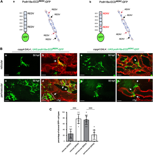
The extracellular domain of Pcdh18a mediates the adhesion of stromal cells to endothelial cells. (Aa,Ab) Fusion proteins expressed in SCPs via the Gal4/UAS system, with the ICD of Pcdh18a substituted by GFP, and tertiary structure prediction for WT (Aa) and mutant (Ab) forms of the ECD, using SWISS-MODEL (expasy.org). Red and black arrows point to two REDV (WT) or AEAV (mutant) motifs in each of two ECDs in homophilic trans interaction. (B) Confocal spinning-disk images of live Tg(cspg4:GAL4; UAS:pcdh18a-ECDREDV or AEAV-GFP; kdrl:ras-mCherry) embryos in the CVP at 50 hpf. (Ba-Bd) Pcdh18a-ECDREDV-GFP expressing reticular (Ba,Bb) or perivascular (Bc,Bd) stromal cells (green) overlaid on endothelial cells (red). Dashed lines indicate the border of each SPvC. (Be-Bh) Pcdh18a-ECDAEAV-GFP expressing reticular (Be,Bf) or perivascular (Bg,Bh) stromal cells (green) overlaid on endothelial cells (red). GFP+ SPvCs typically showed only partial adherence to the endothelium. Arrows indicate GFP+ reticular (Bb,Bf) and perivascular (Bd,Bh) cells, respectively. Scale bars: 10 µm. (C) Histogram showing the percentage of reticular versus perivascular cells among pcdh18a-ECDAEAV-GFP- or pcdh18a-ECDREDV-GFP-expressing stromal cells at 50 hpf. n=10 embryos per condition, from a single experiment (mean±s.d.; ****P<0.0001; unpaired, two-tailed Student's t-test). |